Home » Without Label » Pelvic Anatomy - Medical Illustration Shows The Female Pelvic Anatomy One With Normal Small Bowel Compared With One With Prolapse Of Small Bowel Called Enterocele Royalty Free Cliparts Vectors And Stock Illustration Image 151774867 / • divided into the true and false pelvis by the iliopectineal line.
Pelvic Anatomy - Medical Illustration Shows The Female Pelvic Anatomy One With Normal Small Bowel Compared With One With Prolapse Of Small Bowel Called Enterocele Royalty Free Cliparts Vectors And Stock Illustration Image 151774867 / • divided into the true and false pelvis by the iliopectineal line.
Pelvic Anatomy - Medical Illustration Shows The Female Pelvic Anatomy One With Normal Small Bowel Compared With One With Prolapse Of Small Bowel Called Enterocele Royalty Free Cliparts Vectors And Stock Illustration Image 151774867 / • divided into the true and false pelvis by the iliopectineal line.. Two layers of peritoneum extending from lateral uterus to the…. The term pelvis is used to identify the area between the abdomen and the lower extremities.it can be divided into the greater pelvis and the lesser pelvis. There are four articulations within the pelvis: It is strengthened and supported by several joints and ligaments. Ebraheim's educational animated video describes the anatomy of the pelvis, the bony structures, ligaments, muscles, blood supply, and nerves.this video a.
Sacrum (the large triangular bone at the base of the spine) Pelvic pain can be a sign that there might be a problem with one of the reproductive organs in a woman's pelvic area. The pelvic bones are smaller and narrower. The male pelvis is different from a female's. This quiz is unlabeled so it will test your knowledge on how to identify these structural locations (iliac crest, ischial spine, acetabulum, superior ramus of pubis, posterior superior/inferior iliac spine, lessier.
Pelvic Png Images Pngwing from w7.pngwing.com The pelvic region is the area between the trunk and the lower extremities, or legs. Ebraheim's educational animated video describes the anatomy of the pelvis, the bony structures, ligaments, muscles, blood supply, and nerves.this video a. Each innominate bone is composed of three united bones: It provides attachment to some important muscles in the region, and forms a cavity which accommodates several important internal organs. When you are taking anatomy and physiology you will be required to know the anatomical structure locations of the pelvis. Each is made up of three. Ultrasound uses a transducer that sends out ultrasound waves at a frequency too high to be heard. The pelvis (plural pelves or pelvises) is either the lower part of the trunk of the human body between the abdomen and the thighs (sometimes also called pelvic region of the trunk) or the skeleton embedded in it (sometimes also called bony pelvis, or pelvic skeleton).
The structure of the pelvis supports the contents of the abdomen while also helping to transfer the weight from the spine to the lower limbs.
• pelvis begins at the iliac crests and ends at the symphysis pubis. The structure of the pelvis supports the contents of the abdomen while also helping to transfer the weight from the spine to the lower limbs. Each is made up of three. Female pelvic anatomy what is pelvic pain? Space between bladder and uterus. Visualise your pelvic floor and see exactly what it is, where it's located and why it is important to train this hidden group of muscles. The pelvic bones include the: The pelvic girdle and pelvic spine. Ebraheim's educational animated video describes the anatomy of the pelvis, the bony structures, ligaments, muscles, blood supply, and nerves.this video a. The pelvis is the lower part of the torso. Laparoscopic anatomy of the female pelvic region. Gross anatomy of the pelvis—namely the bladder, uterus, fallopian tubes, ovaries, rectum, and the muscles—has remained unchanged; The pelvis (plural pelves or pelvises) is either the lower part of the trunk of the human body between the abdomen and the thighs (sometimes also called pelvic region of the trunk) or the skeleton embedded in it (sometimes also called bony pelvis, or pelvic skeleton).
The male pelvis is different from a female's. Space between bladder and uterus. Ultrasound uses a transducer that sends out ultrasound waves at a frequency too high to be heard. Each innominate bone is composed of three united bones: However, knowledge of the anatomy of various structures that surround these organs has evolved over time.
Pelvis Anatomy Scheme Royalty Free Vector Image from cdn2.vectorstock.com The main function of the pelvic floor musclesare: Ultrasound uses a transducer that sends out ultrasound waves at a frequency too high to be heard. The pelvis is a basin shaped bony structure formed by the combination of two pelvic bones (hip bones or innominate bones) and the sacrum. There are four articulations within the pelvis: Pelvic anatomy is composed of two innominate (coxal) bones that articulate with the sacrum and proximal. It provides attachment to some important muscles in the region, and forms a cavity which accommodates several important internal organs. The pelvic region is the area between the trunk and the lower extremities, or legs. The pelvis is the lower part of the torso.
Space between bladder and uterus.
The bony pelvis consists of the two hip bones (also known as innominate or pelvic bones), the sacrum and the coccyx. It is strengthened and supported by several joints and ligaments. The pelvis is a musculoskeletal structure that is made up of hip and sacrococcygeal bones, along with several muscular layers. A more detailed account of pelvic anatomy is best found in anatomy texts 1. The pelvic bones include the: The structure of the pelvis supports the contents of the abdomen while also helping to transfer the weight from the spine to the lower limbs. Sacrum (the large triangular bone at the base of the spine) Gross anatomy of the pelvis—namely the bladder, uterus, fallopian tubes, ovaries, rectum, and the muscles—has remained unchanged; A pelvic ultrasound allows quick visualization of the female pelvic organs and structures including the uterus, cervix, vagina, fallopian tubes and ovaries. Ilium, ischium, and pubis, meeting in the acetabular fossa at the triradiate fusion center. Johns hopkins medicine, based in baltimore, maryland Anatomy the pelvis is a ring of bones located at the lower end of the trunk—between the spine and the legs. It is usually divided into two separate anatomic regions:
The structure of the pelvis supports the contents of the abdomen while also helping to transfer the weight from the spine to the lower limbs. The male pelvis is different from a female's. Pelvic anatomy is composed of two innominate (coxal) bones that articulate with the sacrum and proximal. Ebraheim's educational animated video describes the anatomy of the pelvis, the bony structures, ligaments, muscles, blood supply, and nerves.this video a. Pelvic pain can be a sign that there might be a problem with one of the reproductive organs in a woman's pelvic area.
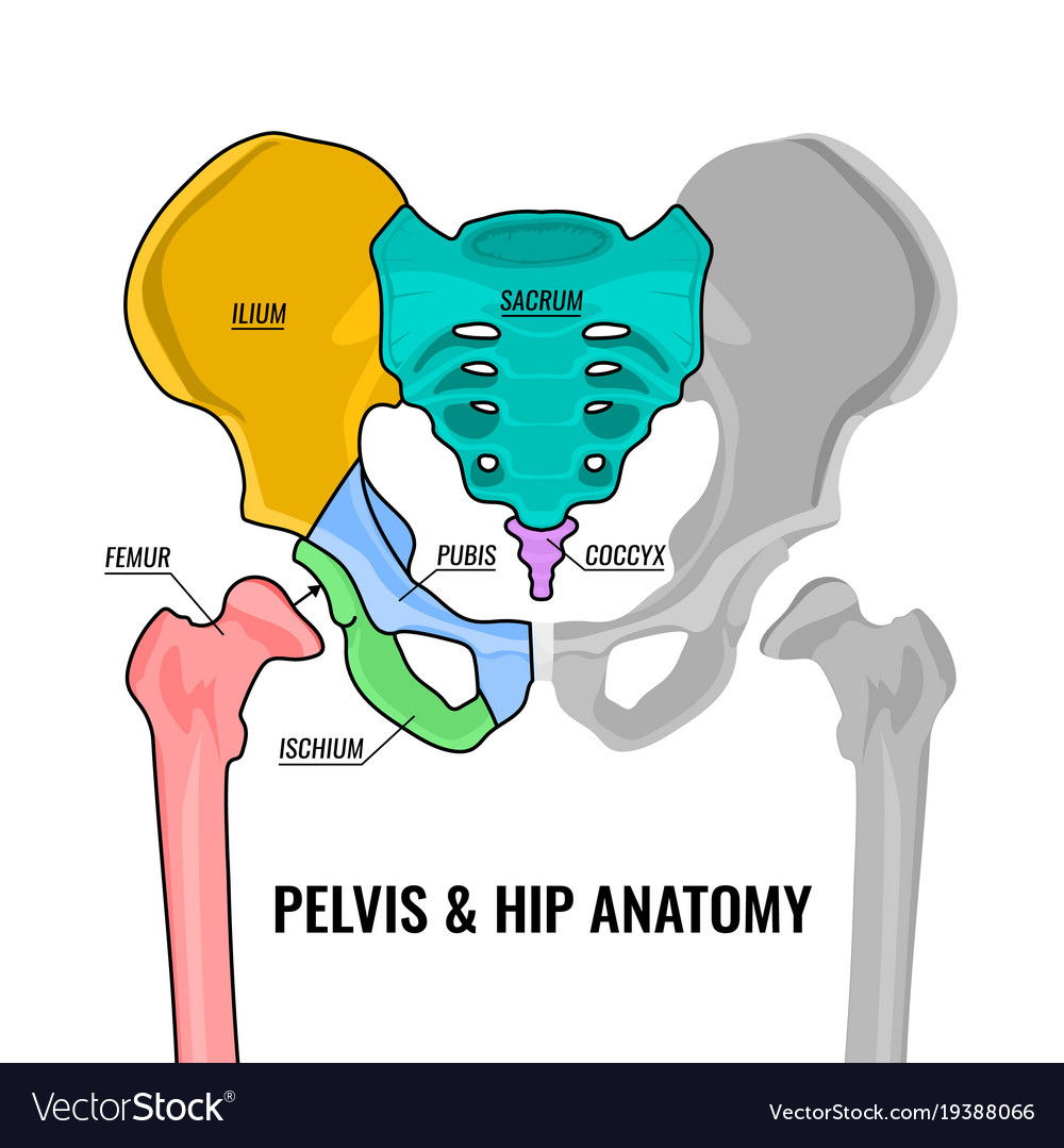
Pelvic Anatomy Unedited Internal Iliac Artery Radical Hysterectomy Ureter And Its Relations Youtube from i.ytimg.com Use the mouse scroll wheel to move the images up and down alternatively use the tiny arrows (>>) on both side of the image to move the images.>>) on both side of the image to move the images. The pelvis consists of the sacrum, the coccyx, the ischium, the ilium, and the pubis. The pelvis is the lower portion of the trunk, located between the abdomen and the lower limbs. The two hip bones (also called coxal bones or os coxae) are together called the pelvic girdle (hip girdle) and serve as the attachment point for each lower limb. Pelvis (hip) anatomy quiz for anatomy and physiology! The anatomy of the pelvis varies depending on whether you are male or female. It's located between the abdomen and the legs. There are four articulations within the pelvis:
The pelvic region is the area between the trunk and the lower extremities, or legs.
The pelvis is a ring structure… Space between bladder and uterus. Pelvic anatomy is composed of two innominate (coxal) bones that articulate with the sacrum and proximal. Pelvis (hip) anatomy quiz for anatomy and physiology! Laparoscopic anatomy of the female pelvic region. Johns hopkins medicine, based in baltimore, maryland There are four articulations within the pelvis: Each innominate bone is composed of three united bones: The pelvis is a musculoskeletal structure that is made up of hip and sacrococcygeal bones, along with several muscular layers. Ebraheim's educational animated video describes the anatomy of the pelvis, the bony structures, ligaments, muscles, blood supply, and nerves.this video a. Anatomy the pelvis is a ring of bones located at the lower end of the trunk—between the spine and the legs. The pelvis consists of the sacrum, the coccyx, the ischium, the ilium, and the pubis. This mri male pelvis axial cross sectional anatomy tool is absolutely free to use.